40 label the transmission electron micrograph of the cell.
a Transmission electron micrograph of leaf cells from E. osiris. Plant ... Labels: Ch, chloroplast; Ne, nucleus; V, vacuole. b Transmission electron micrograph of root cells from E. osiris. Plant was exposed to 0 mg L⁻¹ (control, c, e, g × 30,000) and 15 mg L⁻¹(d ... Label This Transmission Electron Micrograph / Microscopy Innovations ... Label the transmission electron micrograph of the nucleus. Fluorescence microscopy in combination with tem and an ion beam analysis (iba, which allows the evaluation of the chemical elemental distribution) has allowed . Labeling nuclear proteins with electron dense probes in living cells. Label the transmission electron micrograph of the cell.
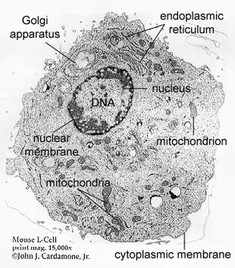
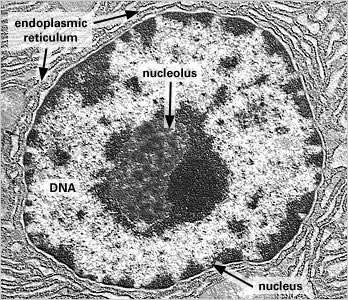
A tour of the cell: View as single page - Open University Figure 2 (a) A transmission electron microscope. (b) A transmission electron micrograph of a frog leukocyte (white blood cell). The nucleus and nucleolus (Section 4.3), mitochondria (Section 4.10) and Golgi apparatus (Section 4.7) can be seen. The dark area of the nucleus contains densely packed DNA.

Label the transmission electron micrograph of the cell.
Plant Cell Nucleus Electron Micrograph : Cell And Organelles Dr Jastrow ... The nucleus (plural = nuclei) figure 7.14 at left a transmission electron micrograph and at right a labeled diagram of a. An electron micrograph of a cell nucleus showing a densely staining nucleolus. Plant cell, electron micrograph 13 plant cells and tissues 29, 30 fiber 11. Electron Micrographs of Cell Organelles | Zoology It is an electron micrograph of endoplasmic reticulum and is characterized by following features (Fig. 11 & 12): (1) It was discovered and named by Porter (1948). (2) It is made up of large number of interconnected and branched tubules, long, flattened and sac-like cisternae and hollow approximately rounded vesicles present all over in the cytoplasm forming a continuous system. [Transmission Electron Micrograph] - 18 images - ebola virus entry into ... [Transmission Electron Micrograph] - 18 images - transmission electron microscopy, brief introduction of transmission electron microscopy authorstream, scanning transmission electron microscopy springerlink, cin2003 ian roberts mast cells in the kidney,
Label the transmission electron micrograph of the cell.. Solved Label the transmission electron micrograph of the | Chegg.com Expert Answer 100% (4 ratings) Explanation - Mitochondrion is filamentous or globular in shape, occur in variable numbers from a few hundred to few thousands in different cells. It … View the full answer Transcribed image text: Label the transmission electron micrograph of the mitochondrion. Labeling the Cell Flashcards | Quizlet Label the transmission electron micrograph of the nucleus. membrane bound organelles golgi apparatus, mitochondrion, lysosome, peroxisome, rough endoplasmic reticulum nonmembrane bound organelles ribosomes, centrosome, proteasomes cytoskeleton includes microfilaments, intermediate filaments, microtubules Identify the highlighted structures Electron Microscopic Study of Cell and Organelles| Important Some of the membranes of endoplasmic reticulum open out in the cell membrane and the others in the nuclear membrane. Golgi apparatus in cytoplasm appears, under an electron microscope, as a pile of 2-layered flattened sacs called cisternae or saccules, lying one within the other.The sacs have flattened or swollen ends near which lie the vesicles of varying size and shape. DP Biology (Cells: 1.1, 1.2, 1.5, 6.3) Quiz - Quizizz The image shows a phagocytic white blood cell as seen with a transmission electron microscope. Which features can be found both within this cell and in a photosynthetic bacterium? DP Biology ... What is the structure labeled X in the electron micrograph of a rat liver cell? answer choices . ribosome. lysosome. mitochondrion. nucleus. Tags: ...
Transmission Electron Microscope (With Diagram) Finally, the electrons are focused by an electromagnetic projector lens (instead of an ocular lens as in a light microscope) on a screen or photographic plate. The final image in a TEM is known as transmission electron micrograph. The salts of some heavy metals, e.g., lead; osmium, tungsten and uranium are often used for staining. Electron Micrographs** Figure 1 Micrograph of a nucleus. 1. Heterochromatin 2. Euchromatin 3. Nucleolus 4. Nucleolar associated chromatin 5. Nuclear envelope Figure 2 Micrograph of a portion of a nucleus: What is the round structure (approximately 3 1/2 inches in diameter) seen in the center of this micrograph? 1. Nucleolar associated chromatin 2. anatomy 10.png - Label the transmission electron micrograph of the ... Label the transmission electron micrograph of the. Study Resources. Main Menu; by School; by Literature Title; by Subject; Textbook Solutions Expert Tutors Earn. Main Menu; Earn Free Access ... Illustrations have been placed from the video to help refresh your memory. List six general examples of cellular processes (several mentioned at the. Q ... Solved Label the transmission electron micrograph of the | Chegg.com Question: Label the transmission electron micrograph of the cell. 0 Nucleus rences Mitochondrion Heterochromatin Peroxisome Vesicle ULAR bumit Click and drag each label into the correct category to indicate whether it pertains to the cytoplasm or the plasma membrane. ICF Contacts the ECF Made of proteins and lipids Surrounds the cell Contains lon channels Organelles
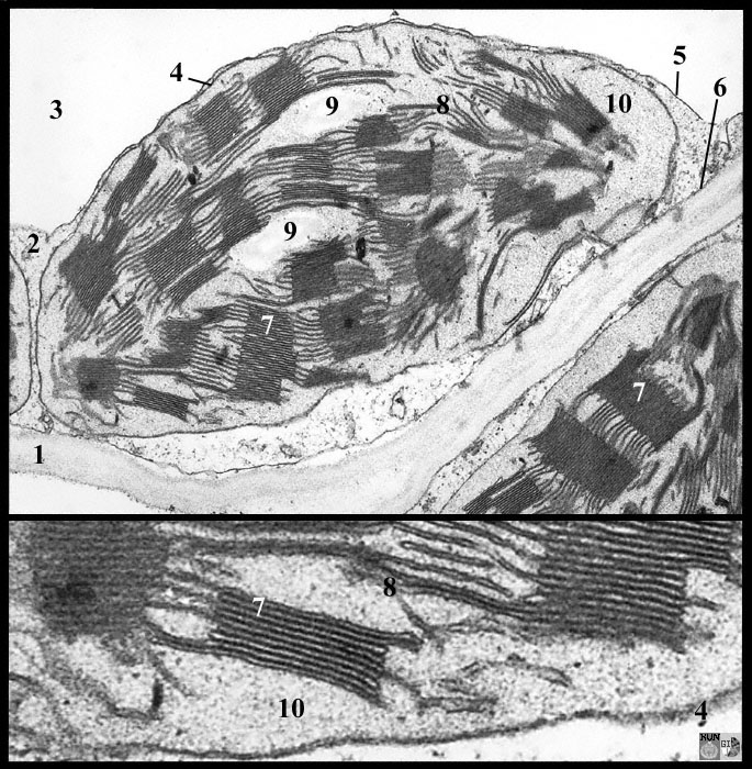
Label the transmission electron micrograph of the cell. 0 Nucleus ... Label the transmission electron micrograph of the cell. 0 Nucleus rences Mitochondrion Heterochromatin Peroxisome Vesicle ULAR bumit Click and drag each label into the correct category to indicate whether it pertains to the cytoplasm or the plasma membrane. ICF Contacts the ECF Made of proteins and... Transmission electron micrograph of cultured mouse mammary cell labeled ... Download scientific diagram | Transmission electron micrograph of cultured mouse mammary cell labeled as shown in Fig. 3; a thick section was used to increase the length of TMV label included in ... Label This Transmission Electron Micrograph : TEM of chloroplast from ... Interpretation of electron micrographs to identify organelles and deduce the functions of specialized cells. Figures label this transmission electron micrograph ( 16, . No microtubule labeling is evident. Label this transmission electron micrograph of relaxed sarcomeres by clicking and dragging the labels to the correct location . PDF Identifying Organelles from an Electron Micrograph The electron micrograph displayed below illustrates many of the plant cell characteristics discussed The cell wall, large central vacuole and chloroplasts are clearly visible Also visible is the clearly defined nucleus containing chromatin Nucleus Chromatin The vacuole in this mature plant cell from a leaf is large, and occupies about 80% of
The Cell: The Histology Guide
Transmission Electron Microscope: Definition, Parts, Working Principle ... In a Transmission electron microscope, the electron beam is transmitted through a very thin specimen or object and forms a highly magnified and detailed image of the sample. This microscope uses electron beams instead of light. The specimen used in Transmission Electron Microscope, should be very thin, less than 100 nm thick.

Smell Receptors Transmission Electron Micrograph Stock Illustration ...
A tour of the cell - OpenLearn - Open University Figure 2 (a) A transmission electron microscope. (b) A transmission electron micrograph of a frog leukocyte (white blood cell). The nucleus and nucleolus (Section 4.3), mitochondria (Section 4.10) and Golgi apparatus (Section 4.7) can be seen. The dark area of the nucleus contains densely packed DNA. Long description.
Topic 1.2 Ultra-Structure of Cells - AMAZING WORLD OF SCIENCE WITH MR ...
The Transmission Electron Microscope | CCBER What is a Transmission Electron Microscope? Transmission electron microscopes (TEM) are microscopes that use a particle beam of electrons to visualize specimens and generate a highly-magnified image. TEMs can magnify objects up to 2 million times. In order to get a better idea of just how small that is, think of how small a cell is.
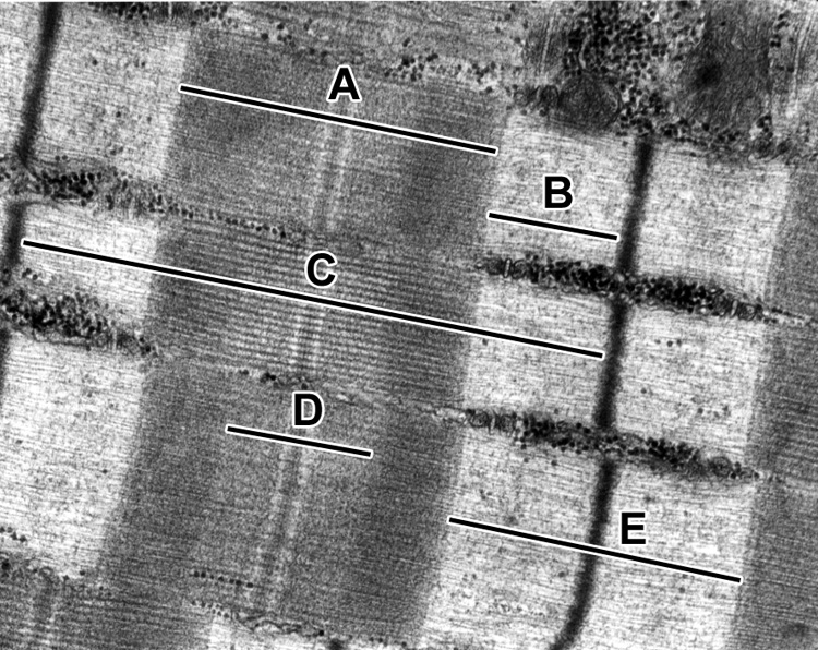
Muscle | histology
Lap Practical #1 EC Flashcards | Quizlet Place the following cytoplasmic structures in the appropriate structural category. Label the transmission electron micrograph of the cell. What would be the consequence if the highlighted structures suddenly became nonpolar? The lipid bilayer would not be able to hold its shape in water and the cell membrane would disassemble.
Basic anatomy
Transmission Electron Microscopy - an overview | ScienceDirect Topics Transmission and scanning electron microscopy have now been used to study the morphology of small intestinal biopsies taken from children as well as adults (Figures 3.26 and 3.27).Certain differences in morphology in children have been observed (Phillips, France and Walker-Smith, 1979).Such studies are now within the scope of routine assessment of mucosal morphology of small intestinal biopsy ...
FluoroNanogold: Fluorescence and Electron Micrographs of Labeled SC35 ...
Transmission Electron Microscope (TEM)- Definition, Principle, Images The working principle of the Transmission Electron Microscope (TEM) is similar to the light microscope. The major difference is that light microscopes use light rays to focus and produce an image while the TEM uses a beam of electrons to focus on the specimen, to produce an image. Electrons have a shorter wavelength in comparison to light which ...
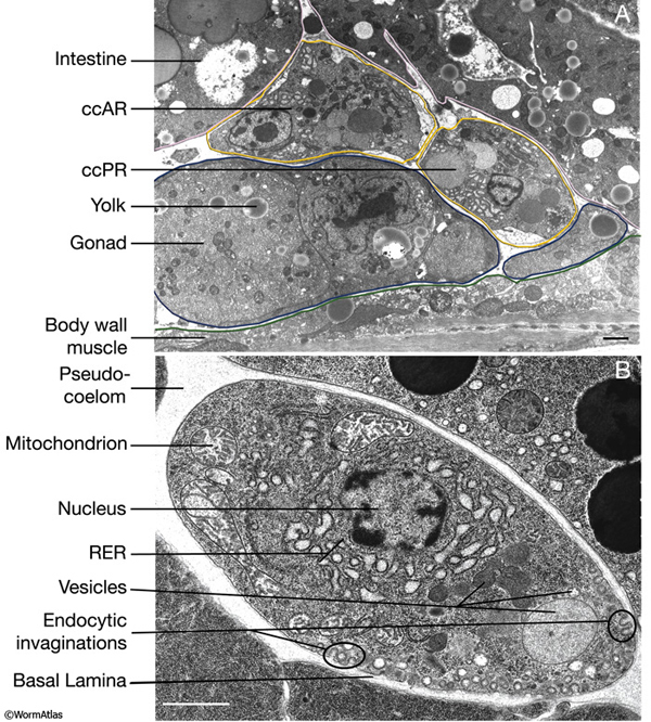
Hermaphrodite Coelomocyte System
Electron Microscopic Structure of a Typical Bacterial Cell The absence of a true cell wall makes these organisms highly plastic and readily deformable, hence mycoplasmas are irregular and variable in shape. The cells may be coccoid, granular, pear- shaped, cluster-like, ring-like or filamentous. These organisms are covered with a unit lipo-protein cytoplasmic membrane, 7.5-10 µm thick.
monograph 1
Transmission electron microscopy DNA sequencing - Wikipedia Workflow of transmission electron microscopy DNA sequencing Step 1 - DNA denaturation As in a standard polymerase chain reaction (PCR), the double stranded DNA molecules to be sequenced must be denatured before the second strand can be synthesized with labeled nucleotides. Step 2 - Heavy atom labeling
Cells Formative Assignment
The Cell | Electron Microscopy | Histology Guide Transmission Electron Microscopy TEM. TEM produces two-dimensional images of a specimen by imaging a thin section with a beam of electrons. Ultrathin tissue sections are stained with heavy metals (such as osmium tetroxide, uranium or lead salts) to enhance contrast. ... Example micrograph of a cell imaged by TEM that explains how these images ...
Post a Comment for "40 label the transmission electron micrograph of the cell."