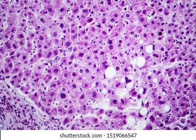
38 label the light micrograph of the liver using the hints provided
All alkaloids in kratom and their action - Reddit The price of the bulk low sodium lemon juice I have been buying spiked from $25/gal to $37/gal delivered, a pretty big increase 1, especially since I'm using about 150mL/day (1 bottle good for about 25 days). Bulk concentrate can also contain a lot of sodium which adds up if using a lot because the label use is extremely small. Module 3 Study Guide ch 24 ch 25Catrina Greene BIO-169-1901.docx Label the parts of the liver and gallbladder using the hints provided. Label the abdominal contents using the hints provided. Label the sagittal section of the mesenteries. Label the mucous membrane tissue from the stomach using the hints if provided. Correctly organize the events of the defecation reflex.
Blood Smear - Understand the Test - Testing.com The blood smear is primarily ordered as a follow-up test when a CBC with differential, performed with an automated blood cell counter, indicates the presence of atypical, abnormal, or immature cells. It may also be performed when a person has signs and symptoms that suggest a condition affecting blood cell production or lifespan.

Label the light micrograph of the liver using the hints provided
Digestive System Flashcards | Quizlet Label the internal features of stomach and duodenum using the hints if provided. Label the biliary passages and associated structures using the hints provided. The liver produces bile and it is stored in the gallbladder until it is released for digestion. Label the abdominal contents using the hints if provided. ... Sets with similar terms Endocrine System and Quiz 5 - histology This electron micrograph shows two of the pituitary cell types in more detail. Two gonadotropes occupy most of the lower half of the figure. Two somatotropes take most of the middle and upper portion of the image. Hormones are synthesized on the rough endoplasmic reticulum (RER) of the two cell types. The Twilight Series is often panned, but at the same time, must have ... Skilled genre writers know that a certain level of artificiality must prevail. It's plot we want and plenty of it. Basically, a guilty pleasure is a fix in the form of a story. The guilty-pleasure label peels off more easily if we recall that the novel itself was once something of a guilty pleasure.
Label the light micrograph of the liver using the hints provided. A&P 2 Lab Practical Final Flashcards - Quizlet Label the anterior view of the larynx based on the hints if provided. Place the following words in order to show the pathway oxygen will diffuse across the respiratory membrane Label the anterior view of the lower respiratory tract based on the hints if provided. Label the photomicrogram of the lung. Which structure is highlighted? (Respiratory) Ileum: Anatomy, histology, composition, functions - Kenhub The small intestine is composed of three distinct parts, the last one being the ileum.At the distal end, the ileum is separated from the large intestine, into which it opens, by the ileocecal valve.The ileum itself is very rich in lymphoid follicles and is attached to the abdominal wall by the mesentery.Its vascular supply is provided by the ileal arteries and its innervation via the coeliac ... The Cell Membrane and Transport | BIO103: Human Biology In contrast, active transport is the movement of substances across the membrane using energy from adenosine ... The tiny black granules in this electron micrograph are secretory vesicles filled with enzymes that will be exported from the cells via exocytosis. LM × 2900. (Micrograph provided by the Regents of University of Michigan Medical ... Parts of a microscope with functions and labeled diagram Q. List down the 18 parts of a Microscope. 1. Ocular Lens (Eye Piece) 2. Diopter Adjustment 3. Head 4. Nose Piece 5. Objective Lens 6. Arm (Carrying Handle) 7. Mechanical Stage 8. Stage Clip 9. Aperture 10. Diaphragm 11. Condenser 12. Coarse Adjustment 13. Fine Adjustment 14. Illuminator (Light Source) 15. Stage Controls 16. Base 17.
Solved Label the light micrograph of the liver using the - Chegg Expert Answer 100% (5 ratings) Transcribed image text: Label the light micrograph of the liver using the hints provided. Interlobular branch of hepatic a 0.19 points Interlobular branch of hepatic portal v Central vein Interlobular connective tissue Print References Bile ductule Interlobular branch of hepatic portal v. Lab 8 : Exercise 38: Digestive System Flashcards | Quizlet Label the light micrograph of the liver using the hints provided Label the abdominal blood vessels using the hints provided Match the following tooth structures with their functions Match the following gastric secretions to the cell that produces it Label the intestinal structures using the hints provided Microscope Parts And Use Worksheet - Google Groups In the field of science, recording observations while performing an experiment is one of the most useful tools available. Apply the mosquito and Charge Equ. The _____ are attached to it. Click country to print this test! This lesson on microscope and label and turn light microscope g stage is playing, define vocabulary and a microscope part. Solved Label the light micrograph of the colon using the | Chegg.com 100% (8 ratings) From above downward: Openin …. View the full answer. Transcribed image text: Label the light micrograph of the colon using the hints provided Muscularis externa Intestinal gland Muscularis mucosae Goblet cell Submucosa Mucosa Opening df intestinal land Reset Zoom. Previous question Next question.
GI Post Lab Flashcards - Quizlet Label the oral cavity and pharynx using the hints if provided. Label the structures of the posterior thoracic wall using the hints if provided. ... Label the light micrograph of the liver using the hints provided. Place the following organs of the digestive tract in order from proximal to distal. 'Label-free' imaging tool tracks nanotubes in cells, blood for ... The nanotubes have a diameter of about 1 nanometer, or roughly the length of 10 hydrogen atoms strung together, making them far too small to be seen with a conventional light microscope. One ... Human Structure 703 HISTOLOGY LAB GUIDE - 2007 Use the key gently to open the top drawer at your place & take out the two small boxes of slides. Place them near the middle of the bench. Do not balance your slide box in the open drawer or leave it close to the edge of the bench. [The sound of a slide box hitting the floor is painful and expensive.]
Dexamethasone Pretreatment Alleviates Isoniazid/Lipopolysaccharide ... Paraffin-embedded liver sections were stained by transferase dUTP nick-end labeling (TUNEL) assay in order to identify apoptotic hepatocytes using TUNEL detection kit (KeyGEN BioTECH, Nanjing, China) following the manufacturer's guidelines. TUNEL-stained liver samples were captured with light microscope (Olympus IX81).
Ileum Histology Slide with Labeled Diagram and ... - AnatomyLearner Ileum Histology Slide with Labeled Diagram and Identification Points 05/11/2021 by anatomylearner The ileum histology slide consists of the four layers like tunica mucosa, submucosa, muscular, and serosa. Here, I will show you the detailed histological features of the wall of the ileum slide with a labeled diagram.
PDF 231 lab survival guide Fall05 - spot.pcc.edu The Microscope (Exercise 3) 1. Learn the care of the miscroscope, as described in your lab manual. Which objective should never be used with oil? 2. Learn the parts of the microscope, such as a. ocular lenses, objectives lenses b. nosepiece, arm, stage c. substage light, iris diaphragm lever, condenser d. coarse adjustment knob, fine adjustment ...
Microscopic Anatomy of the Kidney - Course Hero Trace the flow of fluid/ blood through the renal tubules and the kidney. Describe the glomerular filtration membrane and how it excludes blood cells and proteins from the filtrate. The renal structures that conduct the essential work of the kidney cannot be seen by the naked eye. Only a light or electron microscope can reveal these structures.
Interphase Nuclei of Many Mammalian Cell Types Contain Deep, Dynamic ... EM immunocytochemistry hints at the possibility that a nuclear skeleton may provide tracks for the shuttling ... Samples were examined using either a microscope equipped for epifluorescence illumination and a low light level CCD camera (Axiophot; Zeiss Inc.) or a CLSM. ... Double labeling of tissue sections of normal liver also demonstrated an ...
Pancreas histology: Exocrine & endocrine parts, function - Kenhub Insulin exhibits its effects on most cells of the body, most notably those of the liver, muscles, and adipose tissue. The main function of insulin is related to glucose metabolism, decreasing blood glucose, and sparing proteins and lipids by way of several mechanisms: Stimulates uptake of glucose in insulin-dependent tissues via GLUT4 channels
How to use a Microscope | Microbus Microscope Educational Website The proper way to use a monocular microscope is to look through the eyepiece with one eye and keep the other eye open (this helps avoid eye strain). If you have to close one eye when looking into the microscope, it's ok. Remember, everything is upside down and backwards. When you move the slide to the right, the image goes to the left!
Digestive lab Flashcards | Quizlet Label the mucous membrane tissue from the stomach using the hints if provided. Label the digestive abdominal contents using the hints if provided. Place the appropriate words and descriptions with the picture with the correct highlighted digestive accessory organ. Label the structures of the posterior thoracic wall using the hints if provided.
The Twilight Series is often panned, but at the same time, must have ... Skilled genre writers know that a certain level of artificiality must prevail. It's plot we want and plenty of it. Basically, a guilty pleasure is a fix in the form of a story. The guilty-pleasure label peels off more easily if we recall that the novel itself was once something of a guilty pleasure.
Endocrine System and Quiz 5 - histology This electron micrograph shows two of the pituitary cell types in more detail. Two gonadotropes occupy most of the lower half of the figure. Two somatotropes take most of the middle and upper portion of the image. Hormones are synthesized on the rough endoplasmic reticulum (RER) of the two cell types.
Digestive System Flashcards | Quizlet Label the internal features of stomach and duodenum using the hints if provided. Label the biliary passages and associated structures using the hints provided. The liver produces bile and it is stored in the gallbladder until it is released for digestion. Label the abdominal contents using the hints if provided. ... Sets with similar terms





Post a Comment for "38 label the light micrograph of the liver using the hints provided"